Caries and MIH
Dental caries and molar incisor hypomineralisation
Molar Incisor Hypomineralisation (MIH) is a common developmental condition, defined as a “hypomineralisation of systemic origin of 1-4 permanent first molars, frequently associated with affected incisors”.32 Second primary molars can be similarly affected.33 Molars with hypomineralisation are prone to breakdown. The poor quality of enamel means that they are often sensitive to temperature and sometimes even painful on toothbrushing. These factors combined with increased caries susceptibility can lead to rapidly progressing caries. MIH enamel has an abnormal etching and bonding pattern that compromises restorative outcomes.
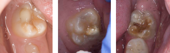
Images show examples of teeth with Molar Incisor Hypomineralisation.
There is a wide spectrum of presentation both in the number of affected teeth and the effect on the teeth. Even within individuals, some teeth will be more affected than others. Lesions range from small, demarcated discoloured areas (white opacities) with no breakdown to large, dark (yellow to dark brown) areas that can fracture off, due to the weakness of hypomineralised enamel, exposing underlying dentine. Patient reported symptoms are variable, and may not necessarily match clinical presentation.
Due to the possibility of rapid post-eruptive breakdown of the enamel (which can be of variable quality), early diagnosis of MIH is key to avoid acute pain and delayed, complicated treatment. If restorations have already been placed, they will often be atypical in shape. This can aid diagnosis of MIH when the lesions are no longer visible.
Assess all hypomineralised molars independently to determine the extent of the disease and likely prognoses. Factors to be taken into consideration when determining whether teeth affected by hypomineralisation are of poor prognosis, include:
- enamel colour in order of severity and increasing likelihood of breakdown: white/cream, then yellow, then brown
- location of the defects in order of severity: smooth surface, then occlusal surface/incisal edge, then cuspal involvement
- sensitivity from brushing or to temperature
- atypically shaped restorations
- any patient reported symptoms