Visual diagnosis
Visual diagnosis
The best method for detecting caries (reducing the risk of under- and over-diagnosis) is visual inspection on clean, dry teeth with good light.
Much clinically relevant information can be gained with an understanding of how dental caries affects the optical properties of enamel and, therefore, why the appearance of caries on the outer surface of enamel differs from caries affecting only the inner border of enamel, at the enamel/dentine junction. Normal healthy enamel is over 98% mineralised and is, therefore, almost transparent. Its apparent colour is due to the colour of whatever lies beneath it, usually healthy dentine.
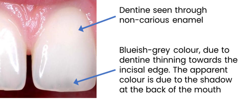
Image shows dentine seen through non-carious enamel and blueish grey enamel at the incisal edge.
Caries affected enamel has a white appearance. Acidic solutions (from cariogenic plaque biofilm, or acid etching solution) preferentially dissolve prism sheaths in enamel, creating pores. These pores refract the light, reflecting it back, instead of letting it pass through.
If the enamel layer is affected, the lesion is matt, opaque, chalky white, as on the cervical region of the mandibular molar below left. When viewing anterior lesions using transmitted light, the lesions will appear dark compared to adjacent healthy enamel due to the light being blocked. Surface enamel lesions with no cavitation are very unlikely to be associated with significant underlying infected dentine and dentinal carious lesions.

Image shows transmitted light being used to examine anterior teeth for carious lesions.
Particularly in a proximal lesion, if caries has reached the enamel dentine junction, the surface layer of enamel may appear unaffected and still transparent. However, the lesion appears opalescent white (like mother of pearl, or translucent plastic), as for the proximal lesion below centre. These tend to be associated with underlying infected dentine and dentinal carious lesions.
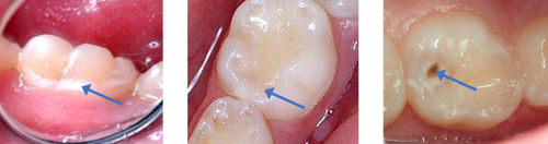
Images show caries on the surface of enamel (left), intact enamel with underlying dentinal caries (centre) and a typical enamel/dentine lesion affecting the distopalatine fissure on a maxilliary molar (right).
The extent of dentinal lesions can be assessed based on the appearance of the overlying enamel. In the example on the right above, the central cavitation is directly visible as dark, carious dentine. This is surrounded by an opalescent white halo where the enamel sub-surface is partially demineralised and reflects light back. Note, the surface of this enamel is not affected, and will appear shiny, rather than the matt white of surface enamel caries. Beyond this, a dark halo is due to direct visualisation of carious dentine through the (as yet) unaffected enamel. Elsewhere in the tooth, healthy dentine is seen through healthy enamel.
If surface enamel pores fill with water (saliva), which has similar optical properties to enamel, then the pores allow light to be transmitted through the enamel. Consequently, teeth must be clean and dry for effective caries diagnosis.
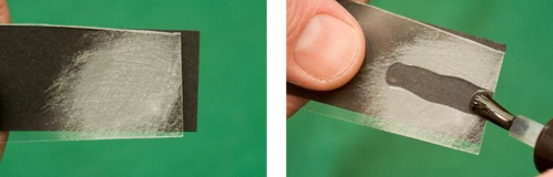
Images show how the abraded surface of a clear acrylic sheet makes it appear white, masking the underlying dark paper (left) and how applying liquid restores the transparency, obscuring the abraded acrylic (right) in the same way that a wet tooth hides enamel caries.
Ensure all teeth are completely clean and dry before assessing for the presence of caries.

Images show mandibular molar before and after cleaning (images on left) and upper permanent teeth before and after cleaning and drying (images on right).
Examine each tooth using a bright, focussed light, and consider using magnification.
- Opalescent enamel adjacent to a stained fissure indicates dentinal involvement.
- A stained pit or fissure without adjacent white opalescent enamel, and with no obvious radiographic signs indicates the carious lesion is confined to the enamel fissure, with no indication for restorative intervention.
- Probing damages pits and fissures and is not an acceptable method for detecting the presence of carious lesions in pits and fissures.
- White opalescent enamel at a marginal ridge indicates a proximal lesion with dentinal involvement. Radiographic examination will confirm the extent of the lesion (see below).
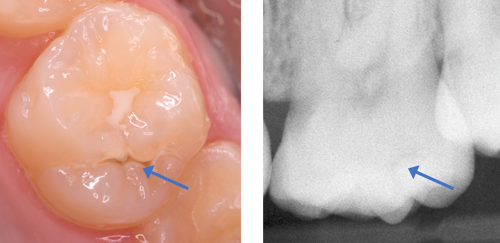
Images show demineralised enamel adjacent to fissure (left) and retinal radiolucency adjacent to the enamel-dentine junction (right).