Assessing risk of progression
Assessing the risk of pain or infection developing before exfoliation
When examining the primary dentition, assess the risk of each carious lesion progressing to pain or infection to decide on the most appropriate management option. Not all carious lesions require operative management. To make this decision consider several factors including:
- extent of the lesion
- site of the lesion
- activity of the lesion
- time to exfoliation
- number of other lesions present in the dentition
- the child’s medical status
- anticipated cooperation of the child, now and in the future
- anticipated cooperation of the parent/carer with the preventive interventions and to attend repeat management appointments
- the range of clinical procedures the clinician has the skill to provide
With so many variables, it is not possible to clearly define specific criteria that will accurately predict which carious lesions will result in pain or infection for the child. The clinician needs to use their skill and judgement when carrying out this risk assessment.
Caries activity is variable, and lesions can arrest or have the potential to arrest. Carious lesions that are slowing or arrested tend to be hard to probing and dark in colour. However, some arrested lesions can be light in colour. Examples of teeth with different carious lesions assessed as at high or low risk of developing pain or infection are shown in the two sets of photographs below. These are intended as a guide only.
Lesions in primary teeth with high risk of causing pain or infection
None of the following lesions have clinically evident signs or symptoms of pain or infection, but are likely to be associated with pain or infection before exfoliation if left unmanaged.
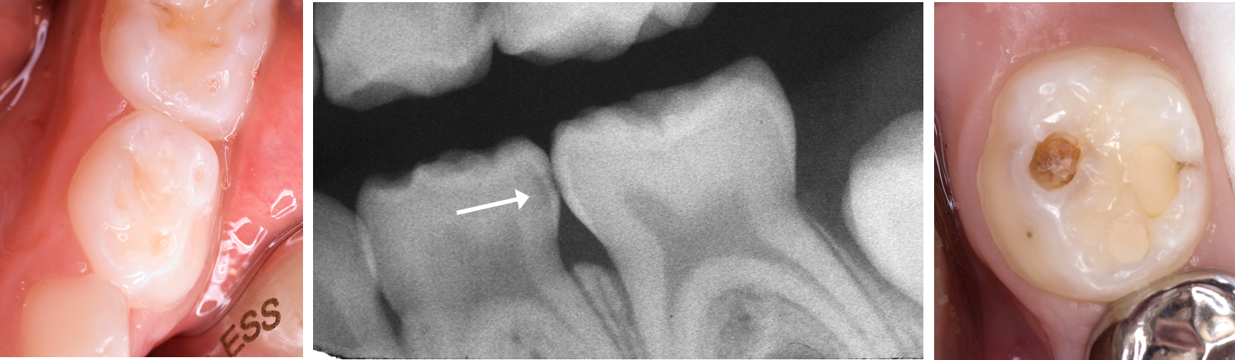
Photographs and radiograph show: initial distal lesion, lower D (left) in a 5-year-old child that is only evident radiographically (centre); cavitated lesion, lower E in a 5-year-old child (right)
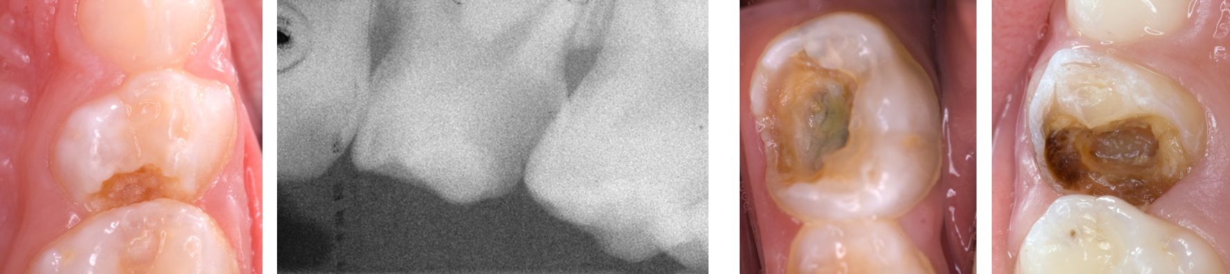
Photographs and radiograph show: upper D with radiographic evidence of pulp exposure (images on left); clinical exposures of necrotic pulps in primary molars and several years before exfoliation (images on right).
Lesions in primary teeth at low risk of causing pain or infection
None of the following lesions has clinically evident signs or symptoms of pain or infection, and, although the teeth do not appear ‘healthy’, it is likely that they will proceed to exfoliation without causing further problems, provided they are closely monitored and the patient is given Enhanced Prevention (see Caries prevention for details).
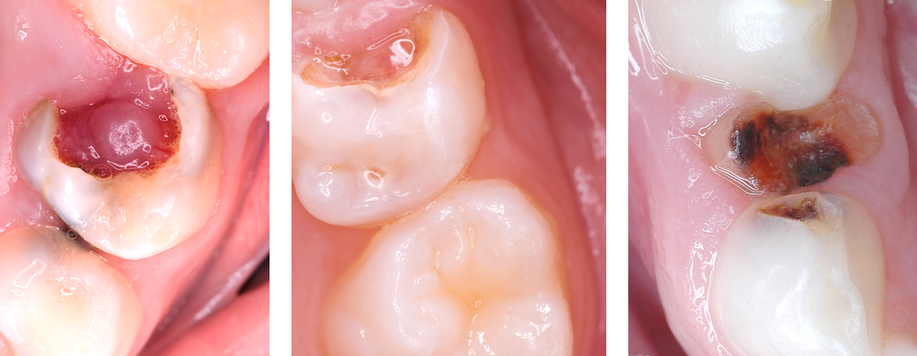
Photographs show: upper Ds and Es (left and centre) with clinical exposures of vital pulps (pulp polyps unlikely to cause infection before exfoliation); retained root, lower D (right; dark coloured and hard).
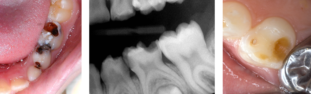
Photographs and radiograph show arrested caries lower CDE (left and centre; dark coloured, hard and cleansable cavity); arrested caries, upper D (right; light coloured and hard).
Note that although a pulp polyp in a carious primary molar indicates that at least one of the root canals is vital, the other canals may be necrotic. If there are signs or symptoms of infection, then extraction or pulp therapy is required.
For each diagnosed carious lesion in a primary tooth, assess the risk of pain or infection developing, prior to exfoliation of the tooth and then decide on a management option.