Permanent teeth - occlusal caries
Permanent teeth with occlusal caries
Initial caries (occlusal)
Description: Visual diagnosis - teeth with noncavitated enamel carious lesions: white spot lesions; discoloured or stained fissures.
Radiographic diagnosis – lesion up to the enamel-dentine junction or not visible.
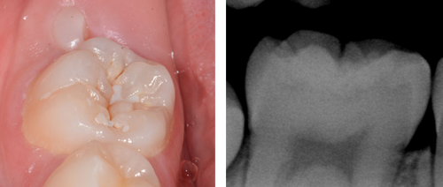
Images show a photograph (left) and radiograph (right) of a permanent tooth with an initial occlusal carious lesion.
Aim: To use a minimally invasive approach to slow or arrest caries and reduce the risk of a permanent molar or premolar requiring an occlusal restoration.
Place a resin fissure sealant (see Sealant/infiltration and Professionally-delivered interventions).
- If initial occlusal dentinal caries is inadvertently sealed in, provided the sealant is maintained, the caries is unlikely to progress.

Image showing a tooth with worn sealant where exposed fissures are now carious. Note that failure to monitor and maintain sealants can allow caries to develop that could have been prevented.
Clinically review sealant for wear and check integrity at every recall visit physically with a probe.
- If the sealant is worn, top it up.
- If the sealant is not adherent to the tooth, remove it and replace.
- If the lesion has progressed, adopt an alternative management strategy.
Radiographically review in line with current recommendations (see Assessing carious lesions).
- If the lesion has progressed into the middle third of dentine, manage as described in Moderate dentinal caries (occlusal) or Extensive dentinal caries (occlusal).
If the tooth is only partially erupted, or the child’s cooperation is insufficient for placement of a resin fissure sealant or a restoration, consider the use of a glass ionomer material as a temporary sealant or restoration (see Professionally-delivered interventions).
Moderate dentinal caries (occlusal)
Description: Visual diagnosis - teeth with enamel cavitation and dentine shadow or a cavity with visible dentine.
Radiographic diagnosis - on a bitewing radiograph these lesions are visible within dentine and may extend into the middle third of dentine (see Assessing carious lesions).
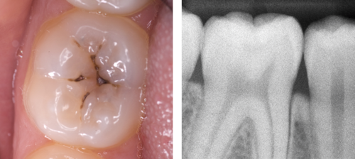
Images show a photograph (left) and radiograph (right) of a permanent tooth with a moderate occlusal dentinal carious lesion.
Aim: To prevent caries progression by placing a sealant or a long-lasting restoration.
If there is no visible dentine, place a resin fissure sealant without removing caries if feasible (see Sealant/infiltration).
Alternatively, carry out selective caries removal or complete caries removal.
- Complete caries removal may be necessary to provide clear walls with enough depth and surface area for a sound restoration. As the caries is not deep there is little risk of pulp exposure with this approach.
Seal the remaining fissures.
Very low certainty evidence suggests sealing moderate occlusal dentinal caries with a sealant material can be as effective as caries removal and restoration. This less invasive approach may be more technically feasible and acceptable in some situations, but it should be noted that regular surveillance of the sealant is required.81
Factors that may influence whether to choose sealant or caries removal include:
- extent of cooperation
- the likelihood of re-attending for active surveillance
- extent of cavitation/lesion
Extensive dentinal caries (occlusal)
Description: Visual diagnosis – teeth with cavitation (this may be extensive) with visible dentine, or widespread dentinal shadow.
Radiographic diagnosis - on a bitewing radiograph, these lesions will extend into the inner third of dentine but there should still be a clear band of dentine that separates the pulp and the carious lesion (see Assessing carious lesions).
Aim: To remove caries, avoiding pulpal exposure and to provide a long-lasting restoration.
Carry out selective caries removal.
Seal the remaining fissures.
If the caries has extended to the pulp, pulp therapy (pulpotomy or pulpectomy/root canal therapy) may be required. The long-term prognosis of the tooth should be considered when treatment planning.