Primary anterior teeth
Primary anterior teeth with carious lesions
Initial caries (anterior)
Description: Visual diagnosis - teeth with white spot lesions/areas of demineralisation confined to the enamel.
Aim: To use a preventive approach to slow or arrest caries and reduce the risk of a tooth requiring a restoration.
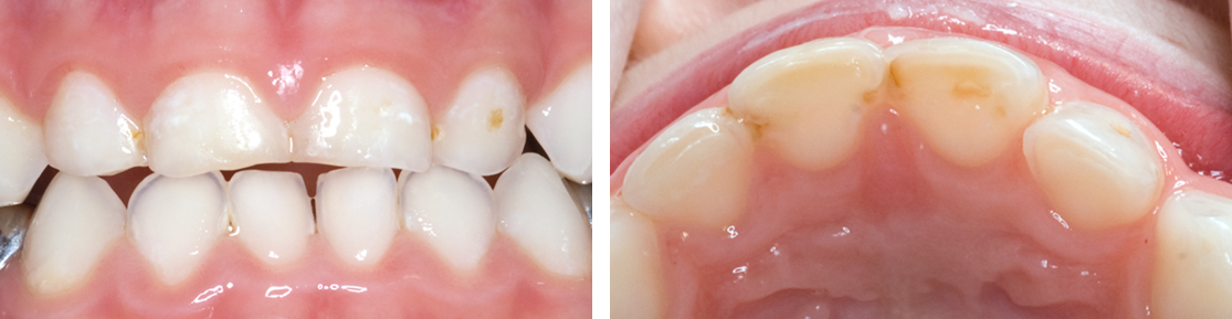
Images show primary teeth with initial anterior carious lesions.
Carry out site-specific prevention.
Monitor at each recall visit and only continue with this approach if caries has arrested and there is no evidence of progression.
If the lesion is progressing, adopt an alternative management strategy.
Some white spot lesions are only detectable on dry enamel, while other more established lesions are visible on wet enamel. Non-invasive management of initial carious lesions is the preferred approach. However, careful active surveillance of enamel lesions is required to enable more intensive prevention or restoration if the lesion is progressing.
Note that if silver diamine fluoride (SDF) is already being used to treat advanced, cavitated lesions in the child, it is acceptable to also apply SDF to initial lesions, with the consent of the child and parent/carer (see Silver diamine fluoride).
Advanced caries (anterior)
Description: Visual diagnosis - teeth with cavitation or dentinal shadow.
Aim: To actively manage caries with minimal use of local anaesthesia and dental handpieces to reduce the risk of causing treatment-induced anxiety.

Image shows primary teeth with advanced carious lesions.
Depending on the cooperation of the child, the preferences of the child and their parent/carer and the extent of the lesion, one of the following approaches may be used.
Carry out selective caries removal and restore using composite, resin modified glass ionomer, compomer, glass ionomer or strip crowns or completely remove caries and restore (see Complete caries removal).
If neither of these approaches is feasible or the tooth is unrestorable, consider non-restorative cavity control, which may include the application of SDF.
The preferred treatment is selective caries removal and restore to avoid iatrogenic damage to the pulp and to reduce the need for local anaesthesia. However, carious lesions which involve the dentine can range from minimal to quite extensive. It may be necessary to remove some tooth substance to provide a good margin on which to place the restoration and to allow placement of adequate bulk of material for stability of the restoration. Care must be taken to detect lesions that have extended into the pulp. In these cases, the tooth will need to be extracted unless a pulp therapy can be carried out.