Primary molars - proximal caries
Primary molar teeth with proximal caries
These lesions are difficult to detect where there is an adjacent tooth. Separation using orthodontic separators can be used but requires an extra appointment. On visible surfaces (such as the distal of E before a first permanent molar erupts or a surface next to where a tooth has been extracted) there might be initial enamel changes, with a white spot lesion only detectable upon drying the enamel. More established white spot lesions will be visible even when the tooth surface is wet. Cavitation or shadowing indicate that the lesion has extended into dentine and is an indication to check radiographically to assess the extent of the lesion and adjacent teeth.
Initial caries (proximal)
Description: Visual diagnosis - teeth with white spot lesions or shadowing.
Radiographic diagnosis - there may be enamel lesions but these do not extend into dentine.
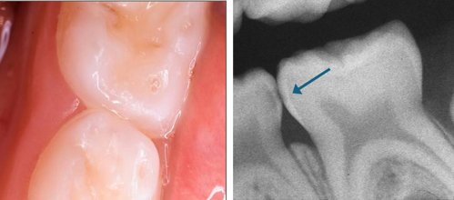
Photograph (left) and radiograph (right) showing a primary tooth with initial proximal carious lesion.
Aim: To use a minimally invasive approach to slow or arrest caries and reduce the risk of a tooth requiring a restoration.
Carry out site-specific prevention and monitor at each recall visit and if the lesion is progressing, adopt an alternative management strategy.
- Only continue with this approach if caries has arrested and there is no evidence of progression.
Alternatively, consider sealing the lesion by placing a sealant or resin infiltration (see Sealant/infiltration) and monitor at each recall visit, repairing and replacing as necessary.
- Active surveillance, maintenance and repair of sealants are essential to avoid the lesion progressing.
If the options above are not suitable, and if silver diamine fluoride (SDF) is already being used to treat advanced, cavitated lesions in the child, it is acceptable to also apply SDF to initial lesions, with the consent of the child and parent/carer (see Silver diamine fluoride).
Advanced caries (proximal)
Description: Visual diagnosis - teeth with enamel cavitation and dentine shadow or a cavity with visible dentine.
Radiographic diagnosis - on a bitewing radiograph, these lesions are visible within dentine and may extend as far as the inner third. There should be a clear band of dentine visible that separates the pulp and the carious lesion (see Assessing carious lesions). Where the radiograph shows there is no clear band of dentine, it is likely that the carious lesion has encroached significantly on the dental pulp and a pulpotomy is necessary (see No clear separation).
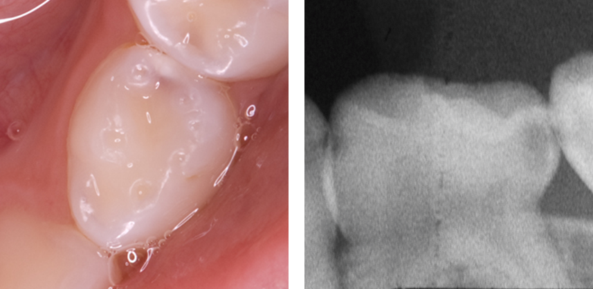
Photograph (left) and radiograph (right) showing a primary tooth with advanced proximal carious lesion.
Aim: To use a minimally invasive restoration to completely seal the carious lesion from the oral environment so that the ecosystem of the plaque biofilm is altered sufficiently to slow or even arrest caries progression.
Without removing the caries, seal in the caries using the Hall Technique.
Alternatively, carry out selective caries removal and restore using composite, resin modified glass ionomer or compomer.
- Where there are symptoms of pain that may be due to food packing or pulpitis with reversible symptoms but the diagnosis is uncertain, a temporary dressing can be placed into the cavity and the patient reviewed 3-7 days later to check symptoms. Resolution of the symptoms at review will indicate that the pulpitis was reversible and a Hall crown or suitable restoration can then be placed. If symptoms do not resolve, or they worsen, then extraction or pulpotomy (see Pulpotomy for primary teeth) should be considered.
If neither of these approaches is feasible or the tooth is unrestorable, consider non-restorative cavity control which may include the application of SDF.
Using the Hall Technique avoids the possibility of iatrogenic damage to the mesial of the first permanent molar from rotary instruments. If restoring, take extreme care (see Avoiding iatrogenic damage).
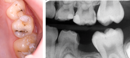
Photograph (left) and radiograph (right) showing a proximal distal dentinal lesion on upper left E.
Complete caries removal should not be carried out on these teeth because of the significantly higher risk of pulp exposure compared with carrying out selective caries removal.
Carious lesions which involve dentine can range from minimal to quite extensive. These lesions can still be managed minimally using a sealing in approach but care must be taken to detect lesions where there is no clear band of dentine visible radiographically that separates the pulp and the lesion. In these cases, the uncertain prognosis should be explained and treatment options discussed (see No clear separation).