Selective caries removal
Selective caries removal and restoration
Suitable for:
- a primary posterior tooth with an advanced occlusal or proximal lesion
- a primary anterior tooth with an advanced lesion
- a permanent tooth with a moderate or extensive occlusal or proximal lesion
- a permanent anterior tooth with an advanced lesion
Aim: To remove sufficient carious tooth tissue to enable an effective marginal seal to be obtained with a bonded adhesive restorative material, inhibiting further progression of residual caries while minimising the risk of iatrogenic pulpal damage.
The evidence that this approach can be effective for both primary and permanent teeth is largely from secondary care and private practice.81
For both primary and permanent teeth, selective caries removal and restoration reduces the risk of pulpal exposure and the time required for cavity preparation.
- Local anaesthesia may not be necessary for primary teeth unless removing sound dentine, but is likely to be required for treating permanent teeth.
- Hand excavation (Atraumatic Restorative Technique, ART) may be useful for cavity preparation in primary teeth.
- Obtaining a marginal seal to arrest caries is essential and dependent on good cavity preparation, which is particularly important for the long-term effectiveness of restorations in permanent teeth.
- For primary molars, the use of plastic adhesive materials is likely to be most successful on occlusal lesions, while a crown placed using the Hall Technique is the preferred option for multi-surface lesions where there can be difficulty achieving a complete peripheral seal.
Technique
Gain access to carious tissue, if necessary using a high-speed handpiece and using local anaesthesia if indicated.
Remove superficial caries with a slow-speed handpiece or excavators, until there is no obvious caries visible at the enamel-dentine junction and the cavity depth allows an adequate thickness of restorative material to be placed.

Image shows a premolar with cavity walls cleared to hard dentine and soft carious tissue remaining on pulpal floor.
Clear the cavity walls to hard (“scratchy”) dentine to provide a good surface for bonding.
- Stained but hard (“scratchy”) dentine may be left unless it causes an aesthetic problem with anterior restorations.
Pulpally, remove enough carious tissue to give adequate depth for a durable restoration, avoiding pulp exposure.
- The consistency of dentine reached pulpally is likely to differ depending on lesion depth. For shallow to moderately deep lesions, caries is likely to be removed until leathery or firm (i.e. feeling of resistance to a hand excavator). For deep lesions, it is likely that some soft dentine caries will be left (i.e. deforming when a hand excavator is pressed on to it and could be easily lifted).
- It is important to avoid iatrogenic damage to adjacent teeth if cutting a multi-surface cavity (see Avoiding iatrogenic damage). Placing a matrix band around the adjacent tooth may help.
- Be aware of the pulp chamber anatomy to reduce the risk of pulpal exposure.
Remove any unsupported or undermined enamel.
Place the restoration, using adhesive material and a bonding system. Do not use conventional glass ionomer materials for restoration of a multi-surface cavity.
Fissure seal unprotected pits and fissures and as many of the restoration margins as possible (see Professionally-delivered interventions).
Monitor for any caries progression using radiographs where appropriate.
Technique for primary incisors
Thoroughly clean the teeth with prophy paste.
- Caries removal will be minimal so local anaesthesia is not required.
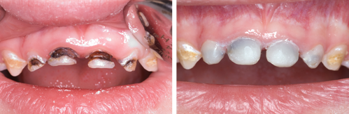
Images show As and Bs managed by selective caries removal and restoration.
Clean the margins of the cavity to ensure that the whole perimeter of the restoration material will be placed on sound tooth substance.
Acid etch the entire crown; wash, dry and apply a bonding system.
Place the composite restoration, either by hand building or using strip crowns.